

General surgeons may not see burn patients every day, but burns are a major public health concern, afflicting more than 1 million people in North America every year. The injuries range from mild to life-threatening, and although they share similarities with any wound a surgeon might treat, they also present unique challenges.
“It’s really important for health care professionals to be able to diagnose and treat burns as quickly as possible,” said Maria Goddard, MD, CWS, a burn and wound care specialist at Goddard Medical, LLC, in Overland Park, Kan.
“Large burns have the potential to create systemic physiological changes in patients; they don’t behave like your typical wound patients because of that systemic inflammatory response,” she noted.
Burn Types and Degree
Burns fall into several categories: thermal (exposure to flame, scalding liquid, contact with hot surfaces), chemical (acid, alkali, organic compounds), electrical (lightning, high voltage, low voltage), and inhalation. Each type has its own concerns and considerations.
“With chemical burns, you’ll want to identify the chemical involved. For instance, patients exposed to hydrochloric acid will have pain out of proportion to the injury, and you’ll want to treat them with calcium gluconate,” Dr. Goddard said, speaking at the 2021 Symposium on Advanced Wound Care virtual spring meeting.
Patients with electrical burns may have hidden injuries and will most likely be seen in an acute care setting, while patients with inhalation injuries need to be checked for carbon monoxide exposure.
First-degree burns, such as sunburns, are superficial and unlikely to need a surgeon’s care. Second-degree burns are subdivided into superficial dermal, which present with blisters and pain, and deep dermal, in which patients might have some diminished sensation due to nerve damage.
“You’ll want to watch second-degree burns closely, as they can go either way—healing on their own or progressing toward coagulation and requiring intervention,” Dr. Goddard said.
Full-thickness third- and fourth-degree burns cause damage to all layers of the skin. “If it goes beyond the subdermis, you may see underlying structures, like bone and muscle,” Dr. Goddard said. “These patients will be at risk for fluid loss and will lose their ability to thermoregulate. We need to monitor these patients for infection as well, which is one of the most common complications.”
Calculating Burn Extent and Fluid Needs
Once you know the mechanism of injury, you’ll want to estimate the total burn surface area (TBSA), and there are a few methods for calculating it. James Howard, MD, a general surgeon in Kansas City, Kan., who is board certified in burn and wound and critical care, favors the rule of nines.
“It’s probably the easiest to remember and is relatively straightforward. Nothing’s 100% accurate, but it’s a good starting guide and will help you with your fluid resuscitation,” he said.
The rule of nines breaks down the body into proportions corresponding to the head, trunk and extremities (e.g., one arm represents 9%). If less than an entire portion is burned, you’d want to take that into account. “Say the burn is only to the anterior portion of the arm, you’d make it 4.5%, or if it’s a smaller area, 2%,” Dr. Goddard said.
Fluid resuscitation comes into the picture when the TBSA is 20% or more. “About 20% is where hypovolemic shock can occur and typically where we would start resuscitation,” Dr. Howard said.
“These patients need a lot of fluid, but the pathophysiology is different than what you’d see with a splenic or liver laceration. You can’t just throw fluid at them, but you need to keep up with their ongoing fluid losses. Sometimes without doing calculations people who don’t often see burn wounds will just give liter after liter, but there really is a ‘Goldilocks spot’ with resuscitation.”
The Parkland formula, 4 mL per hour per percent TBSA, is the calculation most commonly used for determining the volume of lactated Ringer’s needed. “For example, if you have a 20% TBSA, you’ll multiply that by the fluid rate in milliliters and also multiply that by the patient’s weight in kilograms,” Dr. Goddard said.
But this amount may vary a bit. The American Burn Association (ABA) recommends 2 mL per hour for flame burns, 3 mL per hour for pediatric patients and 4 mL per hour for electrical burns.
“The key is to monitor the patient’s urine output and gauge whether you need more or less,” Dr. Goddard said.
Another concern unique to burn patients is their nutritional needs. They may experience a hypermetabolic phase that could last months, even years. “They’ll need greater intake in the acute phase, when they’re the most hypermetabolic. These nutritional needs are reassessed at different stages,” Dr. Goddard said.
Patients who can’t consume adequate nutrition orally may need supplemental nutrition via nasogastric tube, but this raises another problem seen in burn patients: securing any type of tube or line.
“That’s a challenge for every burn center,” Dr. Howard said. “When you can’t secure an IV with tape because the wound is too weepy, suturing is the best and easiest thing to do. Endotracheal tubes can also be difficult—sometimes tying it around the patient’s head with an endotracheal tie is the easiest thing.”
Treatment Details
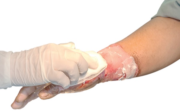
The initial management of a burn wound is a nonlinear cluster of events: removing clothing and jewelry, cooling the wound with warm water (not cold), covering the wound to manage pain and reduce the risk for infection, and optimizing pain control.
“Start with oral medications before you proceed to IV. You’ll want to have frequent discussions with patients regarding their pain level,” Dr. Goddard said.
The mainstay of nonmajor burn care is topical treatment such as silver sulfadiazine (ssD) or mafenide. Prophylactic antibiotics are not recommended.
Dressings are handled as for any wound: Manage exudate, use dressings that reduce infection risk, and consider patient economics and access to care. “Make sure you have adequate pain control to tolerate the changes, and avoid wound cleansers like hydrogen peroxide that could be cytotoxic,” Dr. Goddard said.
Reevaluate within 24 to 48 hours to see if the wound is likely to heal on its own or if excision will be needed.
“For wounds that won’t heal, consider enzymatic debridement before surgery to remove some of the top layer before you need to perform sharp excision. Excision and skin graft placement can be performed in single or multiple steps,” Dr. Goddard said.
“In a patient with a larger TBSA burn where you’ll be limited for donor sites for skin grafting, you’ll want to be more cautious, especially if it’s in the early stages. If your patient can’t go to the operating room, you can use cellular or tissue-based products to aid with the closure of fourth-degree burns,” Dr. Goddard said.
Transfer to Burn Center
Most surgeons would be comfortable managing second-degree burns and doing a skin graft, “say, a 5% or 10% burn on a nonfunctional or noncosmetic area,” Dr. Howard said.
Beyond that, the ABA has identified specific populations of burn patients who should be seen at a burn center: partial-thickness burns greater than 10% TBSA, all full-thickness burns, pediatric burns, inhalation injury, electrical or chemical injury, special anatomic areas (face, hands, joints), burns in combination with trauma, and patients with multiple medical comorbidities.
“Burn wounds are a constant threat to our community, so make sure that even the patients you see in clinic are keeping their water temperatures at appropriate levels and checking their smoke detectors, because we want to protect as many people as possible with prevention,” Dr. Goddard said.