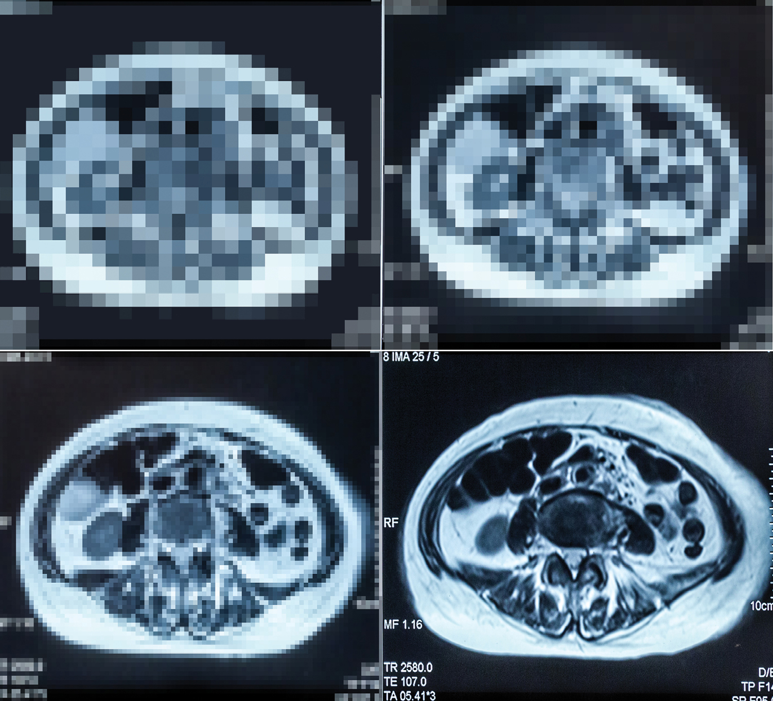
An artificial intelligence algorithm could be used to help surgeons determine which hernia patients have complex cases and are best suited for care at larger referral centers, according to new research.
When presented with pixels from hernia patients’ preoperative CT images, an AI tool developed by investigators at Carolinas Medical Center, in Charlotte, N.C., learned to predict which patients would require component separation or transfer to the ICU because of pulmonary insufficiency, or develop a surgical site infection (ssI), with 64% to 83% accuracy. The work was presented during the Americas Hernia Society virtual annual meeting and received the society’s 2020 Best Paper Award.
“Early studies of AI show promising results aiding surgeons in successful identification of malignancy, ocular and skin pathology, and cerebral bleeding. However, the role of AI in general surgical procedures is unknown,” said lead author and general surgeon Sharbel Elhage, MD. “Determining which hernia patients will require complex surgical techniques or specialized postoperative care remains difficult and is often a subjective decision-making process. The aim of our study was to develop a machine-learning algorithm that could predict complexity and complications in hernia patients based on preoperative CT imaging alone.”
Dr. Elhage’s team pulled in-house data of hernia patients undergoing open abdominal wall reconstruction who had CT scans containing the entire hernia defect. Their outcomes of interest were component separation, defined as transversus abdominis release or external oblique release; pulmonary failure, defined as transfer to the ICU for respiratory complications or intubation; and wound complications, specifically ssIs.
The investigators analyzed images using TeraRecon’s Aquarius iNtuition software and created their tool using the programming language Python and open source Tensorflow and OpenCV frameworks. They standardized all CT images to 150×150 pixels, which was analyzed by an eight-layer convolutional neural network to determine the key image characteristics. Next, they batched the images into training and validation sets.
Overall, the team assessed images from 233 patients. Hernias had an average defect width of 9.4 cm. Patients had a component separation rate of 46.2%, a pulmonary failure rate of 7.3%, and an ssI rate of 22.5%.
The investigators first assessed the AI tool’s ability to predict component separation, Dr. Elhage said. After reviewing CT images 8,000 times while being provided correct information, the algorithm reached 100% accuracy in assessing images in the training set. Next, presented with CT scan images from a validation set of patients, the computer tool accurately predicted whether those patients would require a component separation technique with an accuracy of 74% (P<0.001). Assessing postoperative complications, the tool also reliably predicted patients who would develop pulmonary failure 83% of the time (P<0.001). The AI program also predicted which patients would develop an ssI postoperatively at a rate of 64%, but this did not reach statistical significance (P=0.081).
The work demonstrates that “AI can successfully be used to predict complexity and complications in hernia patients solely based on preoperative CT imaging,” Dr. Elhage said. “This is a groundbreaking proof of concept for the entire surgical field, showing that valuable intra- and postoperative information can be garnered by AI analysis. These tools, once refined, can allow for objective analysis of hernia patients preoperatively, replacing much of the current subjectivity involved in the evaluation and risk stratification of hernia patients, and allowing for the creation of a data-driven, tiered hernia referral system that should improve care for hernia patients around the world.”
Preliminary testing has shown the program has been more successful than a panel of expert hernia surgeons in predicting which patients will require component separation, Dr. Elhage added. The team is continuing work to optimize the algorithms, through methods such as adding additional patient images, assessing what the algorithm is weighting as most important, and adding database variables such as tobacco use, diabetes status and body mass index, with the goal of testing the tool in multicenter prospective trials, he said.
This article is from the March 2021 print issue.


